More than meets the eye
3D printing is helping UC doctors further personalize care, helps patients visualize treatment plans
Today, 3D printing is being used for everything from jewelry creation to producing food.
But the ways in which Dr. Frank Rybicki and the University of Cincinnati Department of Radiology are using the technology are directly impacting the way patients receive care at UC Health, improving their experiences and overall outcomes.
Rybicki, professor in the department, came to UC this year with a decade of medical 3D-printing experience under his belt.
He was part of the team at Brigham and Women’s Hospital and Harvard Medical School, both located in Boston, Massachusetts, that used 3D models for the first U.S. full face transplant in 2011.
"I immediately realized that 3D printing was going to be a game changer for radiology and medicine as a whole,” he says.

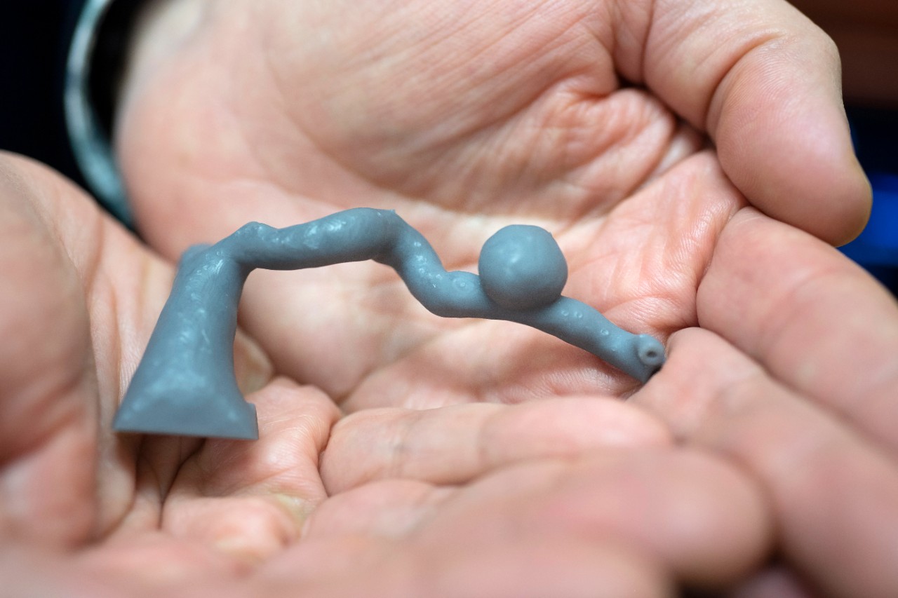
3D model of an artery. Photo credit: Colleen Kelley / UC Creative Services
Using CT, MRI or other forms of medical scans, Rybicki and his team make 3D models of what those scans show, such as tumors or clogged arteries. This then helps physicians make decisions on the best, personalized treatment for the patient as well as explain those plans. These models also help students and residents learn more about anatomy.
The team recently created their first model at UC used for care: a patient’s artery in the liver with an aneurysm.
“We bring the patient’s model directly to the patient,” he says. “It is truly groundbreaking to be able to deliver this personalized care — to show the patient their own disease. This includes tumors, problems from birth or, in the case of blood vessels, blockages. Patient advocacy and personalized medicine are at the center of medical 3D printing. Patients, health care providers and medical organizations strongly embrace the ability for a patient to ‘hold in their hand’ their own anatomy. It increases understanding which promotes communication and the quality of care delivered.”

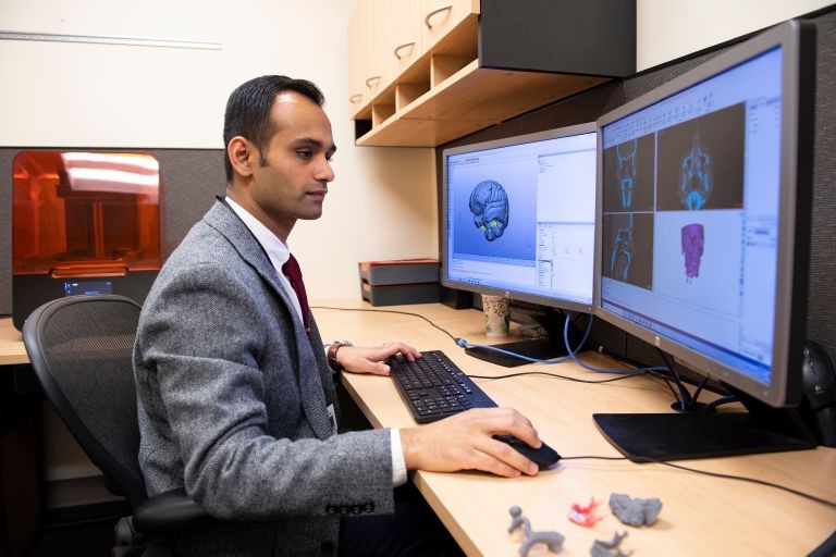
Dr. Arafat Ali, resident physician, assessing scans and inputting images for printing. Photo credit: Colleen Kelley / UC Creative Services
Rybicki is considered a pioneer regarding research on ways to use this technology to help in diagnosis and treatment of patients, in addition to the cost benefit. He’s also working closely with teams at Cincinnati Children’s Hospital Medical Center, which are using the technology to better treat pediatric patients with congenital heart defects.
“It’s an exciting way to meld a technology which is already entrenched in the nonmedical sciences into the medical world,” he says.
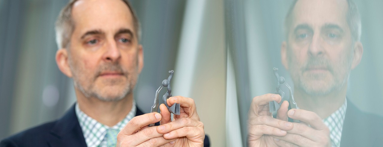
Featured photo at top: Dr. Frank Rybicki by Colleen Kelley/UC Creative Services.
Next Lives Here
The University of Cincinnati is classified as a Research 1 institution by the Carnegie Commission and is ranked in the National Science Foundation's Top-35 public research universities. UC's graduate students and faculty investigate problems and innovate solutions with real-world impact. Next Lives Here.
Related Stories
UC researcher launches app to connect patients to lifesaving...
May 7, 2025
A cardiologist and researcher at UC’s College of Medicine joined the Venture Lab at the 1819 Innovation Hub to launch High Enroll, an app that links clinical trials with willing participants.
Growing heart failure epidemic calls for prioritizing primary...
May 5, 2025
A rapidly growing heart failure epidemic calls for prioritizing primary prevention, according to a new scientific statement from the American Heart Association published in Circulation in April. The statement reviews the current evidence for predicting heart failure risk and offers risk-based strategies for heart failure prevention.
UC students explore transformative trends shaping the future
May 5, 2025
Student researchers at the University of Cincinnati offer speculative looks at the future of healthcare, education and entertainment. We’ll break down their analysis of what to watch for.
