Study uncovers clues to COVID-19 using MRI, CT
A multicenter study, led by UC, finds correlation between the severity of COVID-19 in lungs, brain
Since the pandemic hit, researchers have been uncovering ways COVID-19 impacts other parts of the body, besides the lungs.
Now, for the first time, a visual correlation has been found between the severity of the disease in the lungs using CT scans and the severity of effects on patient’s brains, using MRI scans. This research is published in the American Journal of Neuroradiology. It will be presented at the 59th annual meeting of the American Society of Neuroradiology (ASNR) and has also been selected as a semifinalist for that organization’s Cornelius Dyke Award.
The results show that by looking at lung CT scans of patients diagnosed with COVID-19, physicians may be able to predict just how badly they’ll experience other neurological problems that could show up on brain MRIs, helping improve patient outcomes and identify symptoms for earlier treatment.
CT imaging can detect illness in the lungs better than an MRI, another medical imaging technique. However, MRI can detect many problems in the brain, particularly in COVID-19 patients, that cannot be detected on CT images.
The study was led by Achala Vagal, MD, professor in the department of radiology, and Abdelkader Mahammedi, MD, assistant professor of radiology. Both are UC Health radiologists and members of the UC Gardner Neuroscience Institute.

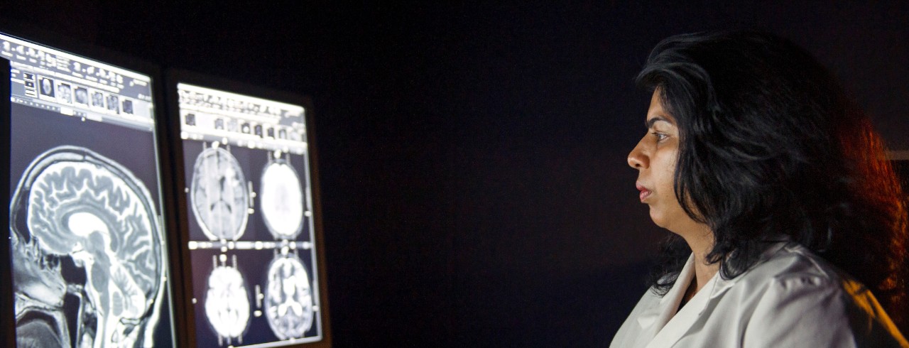
Achala Vagal, MD, author on the study, reviews an MRI scan. Photo/Colleen Kelley
“We’ve seen patients with COVID-19 experience stroke, brain bleeds and other disorders affecting the brain,” says Mahammedi. “So, we’re finding, through patient experiences, that neurological symptoms are correlating to those with more severe respiratory disease; however, little information has been available on identifying potential associations between imaging abnormalities in the brain and lungs in COVID-19 patients.
“Imaging serves as proof for physicians, confirming how an illness is forming and with what severity and helps in making final decisions about a patient’s care.”
In this study, which was conducted not only at UC, but also at large institutions in Spain, Italy and Brazil, researchers reviewed electronic medical records and images of hospitalized COVID-19 patients from March 3 to June 25, 2020. Patients who were diagnosed with COVID-19, experienced neurological issues and who had both lung and brain images available were included.
These results are important because they further show that severe lung disease from COVID-19 could mean serious brain complications, and we have the imaging to help prove it.
Abdelkader Mahammedi, MD

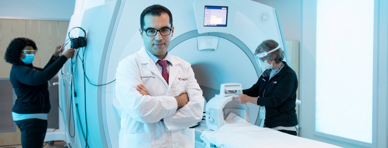
Mahammedi says these findings will help physicians identify potential problems more quickly to implement treatment. Photo/Colleen Kelley
Of 135 COVID-19 patients with abnormal CT lung scans and neurological symptoms, 49, or 36%, were also found to develop abnormal brain scans and were more likely to experience stroke symptoms.
Mahammedi says this study will help physicians classify patients, based on the severity of disease found on their CT scans, into groups more likely to develop brain imaging abnormalities.
He adds that this correlation could be important for implementing therapies, particularly in stroke prevention, to improve outcomes in patients with COVID-19.
“These results are important because they further show that severe lung disease from COVID-19 could mean serious brain complications, and we have the imaging to help prove it,” says Mahammedi. “Future larger studies are needed to help us understand the tie better, but for now, we hope these results can be used to help predict care and ensure that patients have the best outcomes.”
“This also shows the power of collaboration and open science, where physicians and scientists share data," adds Vagal. "This is an international, multicenter study and a heterogenous cohort is always helpful, especially in understanding COVID-19.”
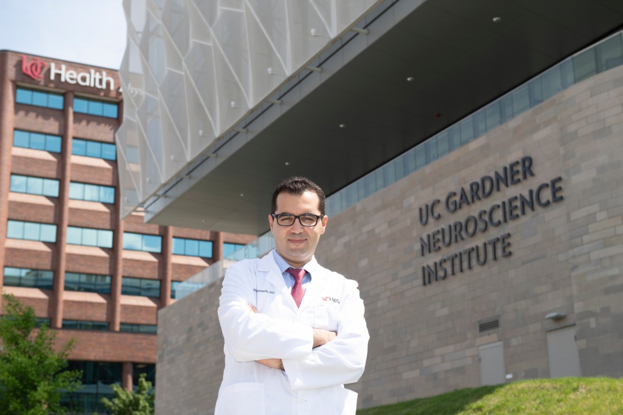
Featured photo of Dr. Abdelkader Mahammedi by Colleen Kelley.
Impact Lives Here
The University of Cincinnati is leading public urban universities into a new era of innovation and impact. Our faculty, staff and students are saving lives, changing outcomes and bending the future in our city's direction. Next Lives Here.
Stay up on all UC's COVID-19 stories, or take a UC virtual visit and begin picturing yourself at an institution that inspires incredible stories.
UC co-authors on the paper include Mary Gaskill-Shipley; Sangita Kapur; Soma Sengupta; Suha Bachir; Lily Wang; Gavin Udstuen; Brady Williamson; Vivek Khandwala; Ram Chadalavada; and Daniel Woo.
This research was supported by the National Institutes of Health (the National Institute of Neurological Disorders and Stroke and National Institute on Aging) (NS103824, NS117643, NS100417, 1U01NS100699, U01NS110772). Researchers cite no conflict of interest.
Related Stories
UC's fourth-year medical students celebrate Match Day
March 24, 2025
At the University of Cincinnati's College of Medicine, 169 students gripped envelopes as they waited to learn their fate on Friday, along with thousands of other medical students across the country. Match Day is the milestone occasion when fourth-year medical students, along with their families, learn where they’ll be spending their residencies.
Add-on metformin promising in ER-positive endometrial cancer
March 24, 2025
The University of Cincinnati Cancer Center's Amanda Jackson was featured in a MedPage Today article commenting on new research that found adding metformin as a combination therapy was tied to deep responses and prolonged progression-free survival in some patients with recurrent estrogen receptor-positive endometrial cancer.
Cancer Center researcher studies unique behavior of pediatric...
March 24, 2025
The University of Cincinnati Cancer Center’s Timothy Phoenix, PhD; Pratiti (Mimi) Bandopadhayay, MBBS, PhD, of Dana-Farber Cancer Institute; and David Jones, PhD, of the German Cancer Research Center have been awarded a $1.2 million grant from the Team Jack Foundation to study pediatric low-grade gliomas.
