55KRC: UC research advances view at the subcellular level

University of Cincinnati cancer biologists have developed a new piece of technology and a new imaging technique that will help researchers glean more detailed data points and see cells in more precise detail when studying the development of cancer and neurodegenerative diseases.
Jiajie Diao, PhD, associate professor in the Department of Cancer Biology in UC’s College of Medicine, told 55KRC's Simply Medicine that a new probe his team developed helps give more details about the cellular environment, such as pH level.
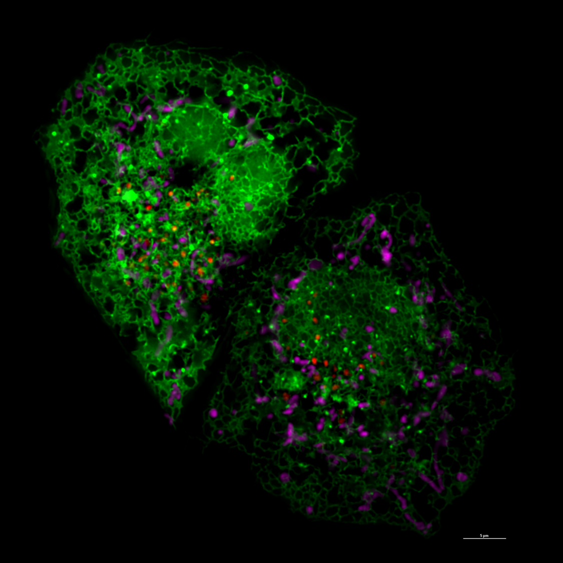
The probe, combined with a new imaging technique that helps measure the shape, distance and location of tiny parts within cells called organelles. Seeing how the organelles interact can provide more information to how these interactions lead to the development of diseases, he said.
"We are actually working with the most advanced image technique to develop new analysis methods, trying to understand and trying to review subtle changes, changes you couldn’t even notice by your eyes," Diao said. "We’re trying to catch diseases at a very early, early stage."
Listen to the Simply Medicine segment. (Note: Segment begins around 23:40 mark.)
Featured photo at top of super resolution image of cell courtesy of Jiajie Diao.
Related Stories
Mural by UC grad honors U.S. military history
July 17, 2024
Local 12 highlighted a new mural by University of Cincinnati graduate and artist Brandon Hawkins that pays tribute to U.S. military history.
Social media fuels extreme political rhetoric
July 17, 2024
UC College of Arts and Sciences Professor Jeffrey Blevins tells Local 12 that online algorithms fuel political polarization on social media.
Camp aims to empower children, teens who stutter
July 17, 2024
A one-week, evidence-based program for children and teens who stutter at the University of Cincinnati will teach kids to communicate effectively, advocate for themselves and develop confidence about their communication abilities. Camp Dream. Speak. Live., which is coming to Cincinnati for the first time July 22-26, began in 2014 at the University of Texas at Austin. The Arthur M. Blank Center for Stuttering Education and Research at UT expects to serve more than 2,000 children at camps across the United States, Africa, Asia and Europe this year.