Research sheds light on minimally invasive neurosurgery approach
UC postdoctoral fellow to present award-winning research at national conference
Brain tumors located in regions that control speech, vision and motor function present additional challenges to neurosurgeons, as damaging the surrounding tissue can cause severe loss of those abilities. Because of this, these regions are known as “eloquent brain areas” and require special attention and approaches to limit damage and deficits.
The University of Cincinnati’s Paolo Palmisciano, MD, was part of a research team that examined how well a minimally invasive approach worked to limit vision and hearing loss in patients following brain tumor surgery.
The research was published in the journal Brain Sciences, and the research team will receive the Mizuho Minimally Invasive Brain Tumor Surgery Award at the American Association of Neurological Surgeons (AANS) annual scientific meeting April 21-24 in Los Angeles.
Technology advances aid surgeons

Paolo Palmisciano, MD, University of Cincinnati postdoctoral fellow.
Palmisciano, a postdoctoral fellow working in the Goodyear Microsurgery Lab in UC’s Department of Neurosurgery in the College of Medicine, contributed to the research prior to his current role at UC. Regardless of the approach a neurosurgeon takes, he explained, advanced technology helps them more specifically navigate directly to the tumor.
These technologies include neuronavigation, which functionally acts as a GPS to guide the surgeon exactly to the tumor. The patient’s MRI is connected with the navigation system, and surgeons can use a pen-like device to register the brain to match the MRI.
“So basically when you do the surgery, you use the pen and you touch the head of the patient and you see on the screen where you are going,” Palmisciano said. “It’s important because otherwise, you can see where the tumor is in the image but you don’t know exactly where in relation to the patient’s anatomy.”
Another technique, called cortical mapping, uses a small amount of electricity to stimulate the brain during the surgery to see if specific regions light up. This helps identify exactly where the regions that control things like speech, motor function and vision are located so that surgeons can avoid these important areas.
“Neurosurgeons know the detailed brain anatomy, but sometimes the tracts are not in the place you think they are because they are displaced by the tumor,” he said. “So you want to do cortical mapping during the surgery to see how far you are from the eloquent area.”
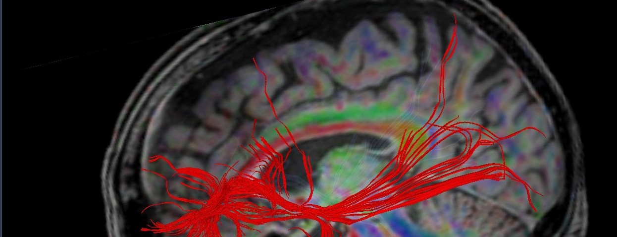
The third technology, tractography, specifically maps the location and direction of white matter within the brain using advanced imaging software. Palmisciano said all three techniques are used together to help surgeons remove as much of the tumor as possible while causing the least amount of damage to the surrounding tissue.
Tubular approach

The research studied the use of a small tube like this one. Once inserted into the brain, neurosurgeons remove tumors through the tube, a more minimally invasive approach. Photo provided by Paolo Palmisciano.
Even using these new technologies, traditional methods of neurosurgery involve removing large portions of the skull, then pulling back large sections of the brain using retractors to get access to the tumor.
"When the neurosurgeon retracts the brain, the brain is mushy and very soft,” Palmisciano said. “If too much traction is used, the surgeon can cause damage to the neurons and also cause tissue death.”
The researchers studied the effectiveness of a different, minimally invasive approach using a small tube. This method involves removing a much smaller portion of the skull, and there is no need to retract the brain.
Since the imaging technology pinpoints where the tumor is located, where to avoid important brain tissue and where the shortest distance to the tumor will be, the tube can be inserted directly into the exact spot of the brain where the tumor is located, then the tumor can be removed through the tube.
“The average length of the tube is 7 centimeters, but you can also use smaller tubes,” Palmisciano said. “The neurosurgeon checks with the neuronavigation that all the tracts are not touched by the tube, puts the tube in and then puts the instrument inside the hole in the tube and to remove the tumor.”
That’s a huge achievement. Instead of doing a big opening, we’re just using this small tube.
Paolo Palmisciano, MD
Study results
The research team analyzed the results of 72 patients who had surgery to remove brain tumors in eloquent areas using the tubular approach from 2018-2021 at the Hospital Infantil Universitario de San Jose in Bogota, Colombia. Because brain tumors in eloquent areas are relatively rare, the prospective observational study was not limited to a specific brain tumor type or region.
“The goal was to see if we can have a lower incidence of complications in this and also to discuss the technique,” said Palmisciano, who contributed to the multicenter analysis of the data.
The research team found that almost 95% of patients in the study had their entire tumors removed using the tubular approach.
“As you can imagine, that’s a huge achievement,” Palmisciano said. “Instead of doing a big opening, we’re just using this small tube.”
Following surgery, 9% of patients had new or worse speech or motor function deficits, which is within the expected complication rate for surgery to remove tumors in these areas of the brain. Palmisciano said a direct comparison to complication rates for more traditional approaches cannot be made with this data since the study included multiple different tumor types.
Going forward, Palmisciano said future studies should look at the effectiveness of the tubular approach for specific tumor types and in more specific regions of the brain to allow for direct comparison.
“Maybe this approach may be of better benefit for particular tumors, so we may be best to use this only for those tumors instead of all types,” he said.
Like with any surgical technique, Palmisciano noted neurosurgeons will not be able to just flip a switch one day and implement tubular approaches if they have not used them before. As more data on its effectiveness is published, it will be important for each surgeon who wants to use a tubular approach to study and practice the technique before using it in an operating room.
Research collaboration
Palmisciano will present the award-winning research on behalf of the research team at AANS on April 24 at 2:45 p.m. during the Rapid Fire Research Forum.
“I feel very grateful to be able to be part of this multi-institutional project, and not for the award itself, but because I think working with different neurosurgeons and experts around the world is always a huge honor for me to learn,” he said. “Neurosurgery is a small field, so having this network helps everyone to move forward in their career.”
Palmisciano will begin a neurosurgery residency at the University of California, Davis in June and said he plans to continue his clinical and academic research in the area of brain tumors.
“I share this award with all the authors, and it embodies both my interest in neuroanatomy and my interest in brain tumors,” he said. “I would consider it as the first real step into academic neurosurgery.”
Other research co-authors and award winners include Nadin J. Abdala-Vargas, Javier G. Patiño-Gomez, Edgar Ordoñez-Rubiano and Hernando A. Cifuentes-Lobelo of the Hospital Infantil Universitario de San Jose in Bogota, Colombia; Giuseppe E. Umana of the Cannizzaro Hospital in Catania, Italy; Gianluca Ferini, Anna Viola and Valentina Zagardo of REM Radioterapia in Vaigrande, Italy; Daniel Casanova-Martinez of the University of Valparaíso in Valparaíso, Chile; Ottavio S. Tomasi of the Paracelsus Private Medical University in Salzburg, Austria; Alvaro Campero of Padilla Hospital in Tucumán, Argentina; and Matias Baldoncini of San Fernando Hospital in Buenos Aires, Argentina.
Next Lives Here
The University of Cincinnati is classified as a Research 1 institution by the Carnegie Commission and is ranked in the National Science Foundation's Top-35 public research universities. UC's medical, graduate and undergraduate students and faculty investigate problems and innovate solutions with real-world impact. Next Lives Here.
Featured photo at top of a MRI brain scan used for tractography techniques. Photo/Wenht/iStock.
Related Stories
Improving treatment for deadly brain tumors
May 5, 2023
The University of Cincinnati's Soma Sengupta was a co-first author on research published in Cell Reports Medicine New that found that a cancer stem cell test can accurately decide more effective treatments and lead to increased survival for patients with glioblastoma, a deadly brain tumor.
Personalized immunotherapy to fight deadly brain tumors
April 13, 2023
The University of Cincinnati is enrolling patients in an Imvax study that will test the effectiveness of a personalized vaccine approach to fight deadly brain tumors.
Using data to improve care for traumatic brain injuries
November 16, 2023
The University of Cincinnati Gardner Neuroscience Institute's Brandon Foreman recently published survey results identifying areas of consensus and needs for further research to develop a standard practice for comprehensively monitoring brain health in the intensive care unit.
