UC-designed probes provide researchers with more detailed data
Collaborative team publishes two new papers that advance cellular, tissue imaging

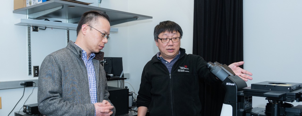
The University of Cincinnati’s Jiajie Diao and Yujie Sun have a simple approach to research: Keep an open mind, and follow the data wherever it leads.
This means even accidental findings are taken seriously, and the path toward their goals is not always straight from point A to point B. This mindset has led to a fruitful collaboration, with the researchers recently publishing articles on new probes that provide more information on how cells function in the journals Chemical Science and Biosensors and Bioelectronics.
Looking deeper into tissue
Previous probes developed by the team focused on a specialized structure called a lysosome that acts as the “recycling center” within cells, reusing broken or malfunctioning materials for different purposes. Abnormal pH levels within lysosomes are associated with cellular malfunctions that can lead to diseases like cancer and Alzheimer’s disease.
Two previous generations of probes provided increasingly more detailed information about lysosome acidity at the cellular level, but the short wavelengths of light that activated the probe were not strong enough to penetrate tissue.
“We had to use shorter wavelengths to excite the probe, and normally it’s visible wavelengths, so their penetration is really limited,” said Diao, PhD, associate professor in the Department of Cancer Biology in UC’s College of Medicine. “We could not go deep, so it’s good for cellular imaging, but it’s not good for tissue or research in living organisms (in vivo).”
The latest advancement from the researchers was to use two lower energy photons with a longer wavelength to activate the probes. Called two-photon imaging, the longer wavelengths can penetrate deeper so that research can be done on animal models and human tissue.
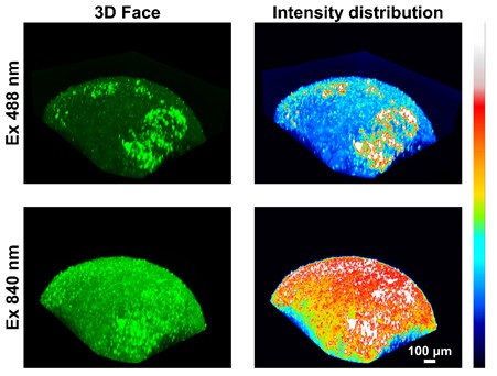
3D reconstruction of microscope images of brain organoids stained with the new three-arm probe are exposed to one-photon (top) or two-photon (bottom) excitation. The two-photon process provides more detail through its fluorescent intensity. Photo provided by Jiajie Diao and Yuije Sun.
Sun and Diao developed three two-photon probes that locate at different areas of cells and can be used to better visualize organelles, or specialized structures within cells.
“It can be used for two-photon microscopy to show 3D images of cells, organoids and tissues,” said Sun, PhD, professor in UC’s Department of Chemistry. “We are developing new two-photon-absorbing fluorescent probes which are sensitive to various factors, such as pH, viscosity and ionic species.”
“Many times, tissue or even in vivo measurement is more important than cellular,” Diao added. “This probe will illuminate individual organelles, and it’s much better than the commercial two-photon probes currently available.”
Detailing individual cell viability
Researchers testing new treatments for diseases like cancer measure cell viability, or whether or not a cell is dying or remains active, after coming into contact with a treatment.
“When you give a treatment, the first thing you always check is the cell viability,” Diao said.
Traditionally, cell viability was measured by looking at a large population of millions of cells at a time, which Diao said is like giving an entire group of students the same exam grade based on their average score.
“As everyone can feel at some time, it’s unfair, because everyone is different,” Diao said. “And every cell is different.”
Progress in the field of cell biology has led to new interest in measuring individual cell viability, which is like grading each student based on their own performance on the exam. This provides more specific insights to researchers by showing how different types of cells react to treatments being tested.
When working on the previous probe that measured cells’ acidity levels, Diao and Sun created a control version that was not sensitive to changes in pH levels. Completely by accident, they found that even though this version of the probe was structurally similar, it behaved differently, locating itself at different organelles.
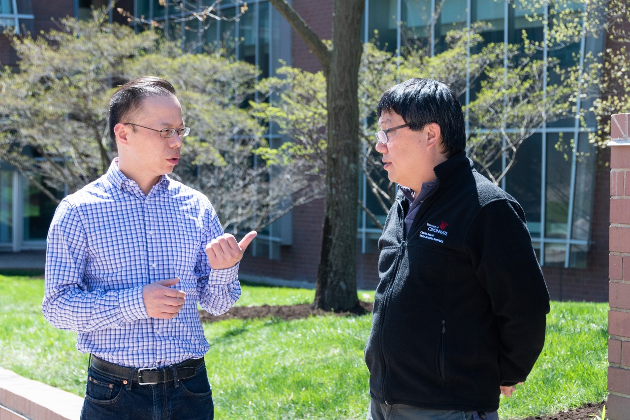
Yujie Sun, left, and Jiajie Diao have a conversation outside of the Vontz Center. Photo/University of Cincinnati Cancer Center.
The new probe initially stains the cell’s mitochondria, which acts as the power plant providing energy to the cell. But when the cell is damaged, the probe moves itself and stains the nucleus, which contains the cell’s genetic material.
“Eventually the signal on the mitochondria will start getting dim, and the signal on the nucleus will get stronger, so by measuring the color intensity ratio between the mitochondria and the nucleus, we can quantitatively assess the viability of individual cells,” Diao said. “That’s a very new concept. Nobody has done this before.”
Long term, the team hopes the probe can be used to learn more about biological differences that affect whether a cell is immediately killed by a treatment or develops resistance and is unaffected.
“There must be something different,” Diao said. “We want to kill the bad cells and we want to make the good cells live longer. We want to assist that at an individual cell level.”
Sun said they will also work to extend what is known as the probe’s spectral window, or the maximum wavelength that can be used to activate the probe.
“Because longer wavelength photons have better tissue penetration, we will be able to see deeper,” Sun said.
I think the most important thing is always keeping an open mind, communicating frequently and not getting limited,. We always say data is data.
Jiajie Diao, PhD
Successful collaboration
Sun and Diao are the co-directors of UC’s Center for Chemical Imaging in Biomedicine, which has an aim of pushing the boundaries of imaging through developing new methods, probes and equipment.
Since joining forces, the duo has been prolific in publishing research and making advancements. Diao credits their joint mindset of not having any preconceived notions of where the research may lead for their success.
“I think the most important thing is always keeping an open mind, communicating frequently and not getting limited,” he said. “We always say data is data. Many times the biggest fight will be the fight between some preset imagination.”
With larger goals always in mind, the research team constantly adjusts expectations and goals when they see an opportunity for tangible progress.
Diao noted they originally started working together with the aim of immediately developing a two-photon probe, but they didn’t have the right experience. Rather than toiling away aimlessly, they switched gears, developed cellular probes and gained the knowledge needed to create the two-photon probes.
Sun said another key aspect of the partnership is chemistry graduate student Rui Chen, who Diao and Sun co-advise.
“Rui has been the bridge between our groups, and therefore Jiajie is fully aware of the progress we are making in my lab,” Sun said. “The complementary expertise between our two groups really makes us a great team to work synergistically together. I’m optimistic about our future achievements in bioimaging.”
Next Lives Here
The University of Cincinnati is classified as a Research 1 institution by the Carnegie Commission and is ranked in the National Science Foundation's Top-35 public research universities. UC's medical, graduate and undergraduate students and faculty investigate problems and innovate solutions with real-world impact. Next Lives Here.
Featured photo at top of Sun, left, and Diao, right in the laboratory. Photo/University of Cincinnati Cancer Center.
Related Stories
Cincinnati researchers want to know if MRIs can work better
June 28, 2024
WVXU and the Cincinnati Business Courier highlighted a new collaboration between the University of Cincinnati College of Medicine, UC Health GE HealthCare, JobsOhio, REDI Cincinnati and Cincinnati Children’s to create an MRI Research and Development Center of Excellence located on UC’s medical campus.
UC opens Blood Cancer Healing Center
June 28, 2024
Media outlets including WLWT, Local 12, Spectrum News, the Cincinnati Enquirer and Cleveland.com highlighted the opening of the University of Cincinnati Cancer Center's Blood Cancer Healing Center.
New project aims to better support teen mothers in Adams County
June 27, 2024
WCPO highlighted a partnership between the University of Cincinnati and the Adams County Health Department that is aiming to provide better support for teen mothers in the county.
Free Wi-Fi, work area coming to Greater Cincinnati
June 27, 2024
St. Lawrence Park in Price Hill now has free Wi-Fi and a furnished outdoor space for community members to access digital needs. The space is part of The Nodes Project, which stands for “Neighborhoods of Design Engagement": a collaboration between UC DAAP communication designers and community entities.
Study aimed at reducing opioid overdose deaths presents results
June 27, 2024
The University of Cincinnati's John Winhusen and Caroline Freiermuth discussed the evidence-based practices implemented during the HEALing Communities Study to fight the opioid epidemic with Spectrum News.
UC alum credits journalism program with early success
June 26, 2024
Zachary Jarrell came to the University of Cincinnati in 2019 to pursue a degree in statistics. In 2023, he graduated with a Bachelor’s in Journalism. For many undergraduates, the journey through college rarely takes the expected track. Detours happen, and majors change. When plans switch up, it can be helpful to a student’s success to find support. For Jarrell, it was the people he worked alongside in the journalism department who helped him on his journey. It has left a lasting impression on his life so far, guiding him to multiple internships as an undergraduate, real-world experience in prominent news outlets, and eventually a successful career in the highly competitive field of journalism.
University of Cincinnati, UC Health collaborate with GE HealthCare on MRI research center
June 26, 2024
The University of Cincinnati and UC Health are collaborating with GE HealthCare, JobsOhio, REDI Cincinnati and Cincinnati Children’s to create an MRI Research and Development Center of Excellence located on UC’s medical campus.
The health impact of living near a natural gas leak
June 25, 2024
UC College of Arts and Sciences Professor Amy Townsend-Small talks to the BBC about the health issues faced by neighbors of leaking natural gas wells.
Male birth control gel inches towards FDA approval
June 25, 2024
The University of Cincinnati's Wesley Baas spoke with Yahoo News and the Cincinnati Enquirer about a new male birth control gel that could be close to receiving FDA approval.
University of Cincinnati Cancer Center launches Blood Cancer Healing Center
June 25, 2024
The University of Cincinnati Cancer Center celebrated the opening of its state-of-the-art Blood Cancer Healing Center June 24.