UC engineering student works to advance ultrasound technology
Elmira Ghahramani's research focuses on liver tumor ablation
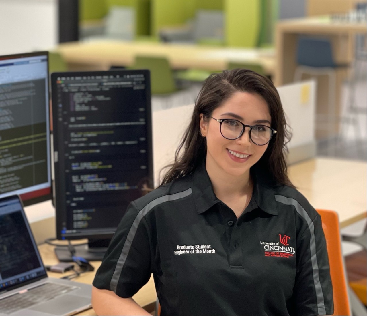
Elmira Ghahramani, a biomedical engineering PhD student at the University of Cincinnati, first found an interest in medical imaging during her undergraduate studies in Tehran, Iran. Her research aims to address limitations and enhance the efficacy of ultrasound imaging, specifically as it applies to liver tumor thermal ablation. She collaborates with professionals from Cincinnati Children's Hospital and was named Graduate Student Engineer of the Month from the College of Engineering and Applied Science.
Why did you come to UC to study?
Among the graduate schools I was considering for enrollment, the University of Cincinnati stood out for its active and strong biomedical ultrasound imaging group. UC's multiple labs are actively engaged in this field, guided by renowned professors, and equipped with state-of-the-art ultrasound imaging instruments that provide an ideal setting for advanced research. The collaborative environment between health care professionals and engineering researchers, coupled with the labs' strategic location at the core of the medical campus significantly influenced my decision to select UC for my doctoral studies.
Why did you choose your field of study?

Elmira Ghahramani
Biomedical imaging offers a unique blend of technology and significant real-world applications. Through our multidisciplinary research, I have used my engineering skills to fill in some clinical gaps to help improve healthcare for patients with cancer. Furthermore, ultrasound imaging is non-invasive, safe, affordable, and widely used in medicine with broad potential, including real-time 2D/3D imaging and characterization of the mechanical properties of the tissue. This imaging modality is a pivotal tool for diagnosis, monitoring, and even treatment. Therefore, as healthcare continues to evolve, there is a growing need for professionals who can develop and refine ultrasound technologies, improving their accuracy and versatility.
Briefly describe your research work? Why does it interest you?
My Ph.D. work aims to enhance the efficacy of ultrasound imaging in the thermal ablation or removal of liver tumors. At the Biomedical Acoustics Laboratory, we do this using our innovative method, 3D ultrasound echo decorrelation imaging.
This is a computationally efficient method that spatially maps heat-induced changes in ultrasound echoes over millisecond time scales and can provide feedback on ablation progress while the tumor is being ablated to avoid over or under treatment.
We explored advanced image and signal processing methods, including real-time feedback control algorithm, temperature prediction, registration among various imaging modalities and more. Using National Institute of Health funding, we have evaluated our method ex vivo (outside the body) in bovine and human liver and in vivo (inside the body) in swine liver, as well as in the percutaneous ablation of human liver tumor. This was done in collaboration with the Department of Radiology at UC. It is quite interesting to me that by developing novel monitoring methodologies and tools, we can enhance the accuracy, precision, and safety of tumor ablation procedures while minimizing the risk to the surrounding healthy tissues.
What are some of the most impactful experiences during your time at UC?
Through my academic coursework and hands-on experience, I have gained a solid foundation in ultrasound imaging techniques, signal and image processing, and software development. I have also had the privilege of working alongside esteemed researchers and clinicians, witnessing firsthand the challenges faced in liver thermal ablation procedures and the critical role ultrasound imaging can play in targeting and ensuring effective treatment outcomes.
What are you most proud of accomplishing?
I took the initiative to extend 3D echo decorrelation imaging to include advanced artificial intelligence techniques to improve the delineation of the ablation zone and temperature prediction for monitoring thermal ablation. For this purpose, I spent a few months earning certificates in the online machine and deep learning courses. I employed my knowledge to augment our echo decorrelation imaging technique, the result of which I presented at the recent Acoustical Society of America international conference in Chicago in May 2023, with the proceedings paper ready to be published. This work led to a new collaboration with the researchers from the Cincinnati Children's Hospital Medical Center and a postdoctoral opportunity at a strong ultrasound research laboratory at Cornell University.
What are your plans after earning your degree?
I will move to New York City to start my work as a postdoctoral researcher at Weill Medical College of Cornell University.
Outside of your research, do you have any other hobbies?
I do have a few other activities and hobbies that I enjoy outside of my professional life. I am passionate about oil painting and find it a therapeutic and creative outlet. I also love to write poetry, which I think is the most beautiful form of self-expression. I also run four or five days a week after work.
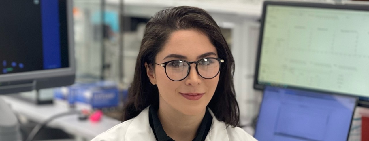
Featured image at top: Elmira Ghahramani in the lab. Photo/provided.
Related Stories
How to prevent concussions in football? Better helmets
March 6, 2023
Football helmets made by four leading manufacturers showed vulnerabilities in tests designed to better understand player concussions, according to a study by the University of CIncinnati.
Engineering students help track pandemic in Ohio
January 10, 2024
The University of Cincinnati has led a three-year project to monitor COVID-19 in wastewater for the Ohio Department of Health. The surveillance project has helped health officials make early interventions to prevent sick students from spreading the virus across campus.
UC engineering student works to improve speech science technology
November 9, 2023
Speech is one of the most complex tasks many people perform in their daily lives. University of Cincinnati graduate student, Sarah Li, is applying principles of biomedical engineering to the study of speech. She has received several awards for this research and was named Graduate Student Engineer of the Month from UC's College of Engineering and Applied Science.
