UC engineering professor awarded $2M NIH grant
Engineering professor Leyla Esfandiari is studying exosomes for potential nerve regeneration

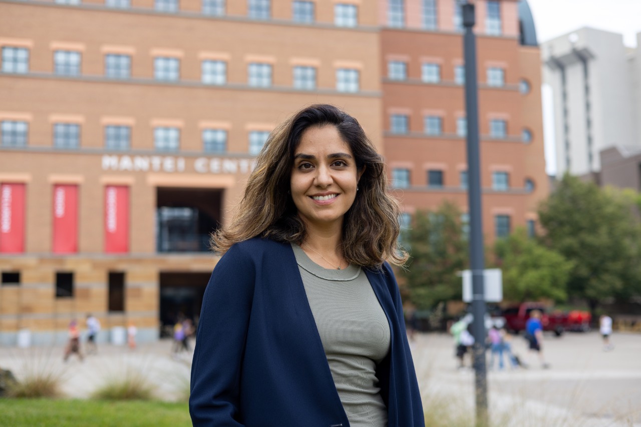
Leyla Esfandiari, an associate professor of biomedical engineering at the University of Cincinnati, has received $2 million from the National Institute of General Medical Sciences to fund her research on small extracellular vesicles called exosomes and the role they play in nerve regeneration as treatment for nerve injuries or other neurodegenerative diseases.
Esfandiari has been studying exosomes for several years. In 2018, her team developed a new lab-on-a-chip device that uses circulating exosomes for noninvasive point-of-care screening. The device extracts exosomes from biofluids in an expedited and cost-effective manner and it was granted a patent in January 2023. That project was partially funded by the National Science Foundation CAREER grant in 2021.
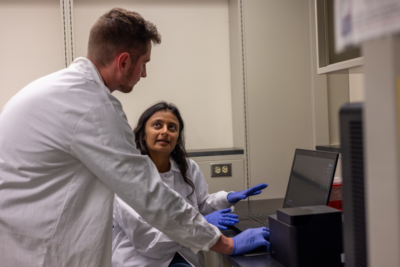
UC students Maulee Sheth and Maksym Krutko work with a nanoimager in Leyla Esfandiari's lab. Photo: Corrie Mayer/CEAS Marketing.
"Previously, we made the device that can help to purify exosomes from biofluids like blood or saliva. We can now extract and purify these vesicles that are secreted from any type of cells and further utilize them for either diagnostic or therapeutic applications," Esfandiari said. "This NIH award is a great opportunity for us to start a new venture on investigating the role of exosomes as new therapeutics for nerve regeneration. We are combining the technologies that we developed in our lab to tackle a new problem in medicine, which is regeneration."
The study of extracellular vesicles is a relatively new field as they were discovered only 40 years ago. At first, they were seen as the garbage bin of cells, simply cycling out the unwanted information. However, once scientists dug deeper into these small vesicles, they quickly realized their impact as carriers for circulating biomarkers.
It is now known that exosomes are important vesicles for cell-to-cell communication. They circulate in the body, and contain messages through micro-RNAs, proteins and lipids. When they are released by cells, they travel through biofluids or pass through tissue and can pass their contents into the recipient cell, therefore changing its genetic coding or function.
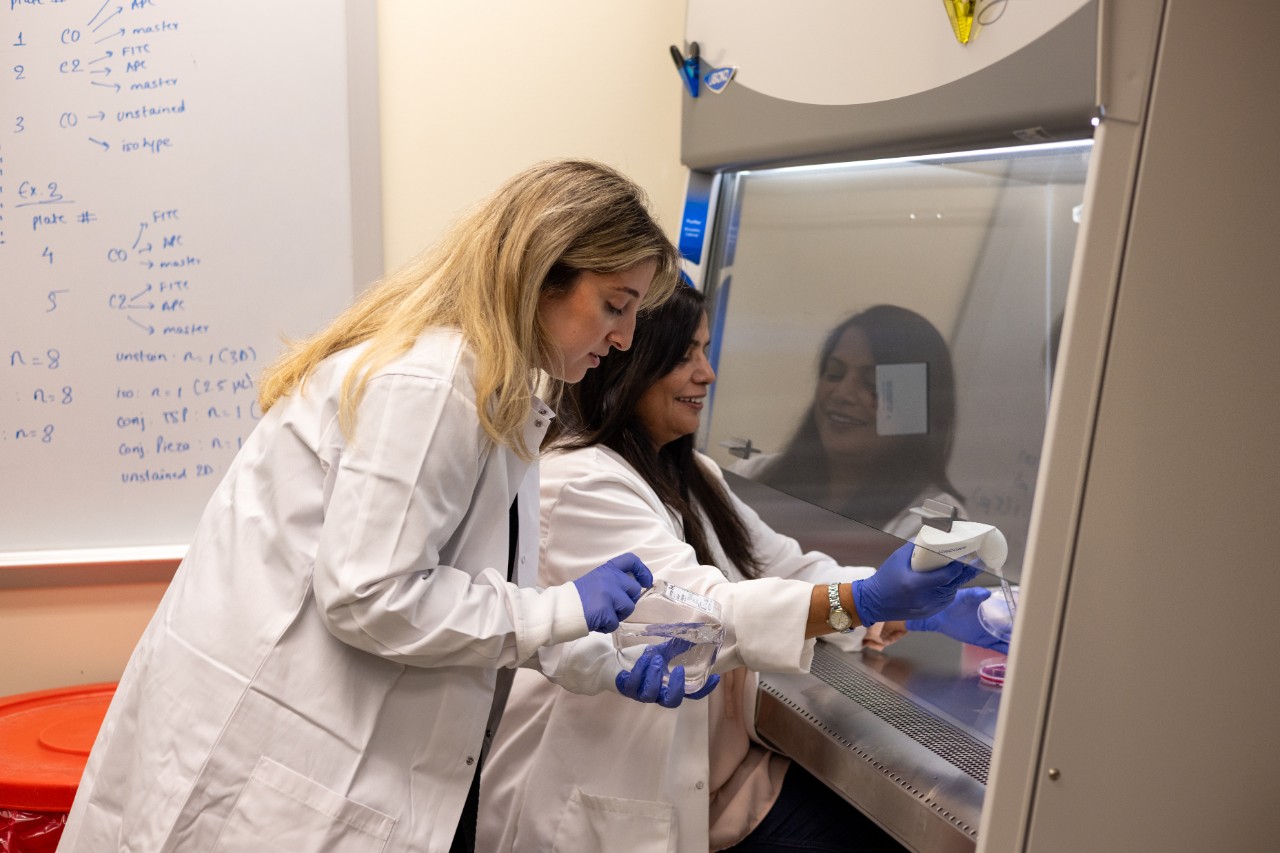
Yara Izhiman and Manju Sharma work on extracting exosomes from patient samples. Photo/Corrie Mayer/CEAS Marketing
The team is researching whether these vesicles will have a different genetic coding once electric fields are applied to their parental cells, thus having the ability to pass the new coding on to its neighboring cells. More specifically, if the micro-RNAs are helping with the regeneration of neurons, they could potentially be used in treatment for nerve injury or neurodegenerative diseases.
The inspiration for this work came from Esfandiari's existing knowledge about the field of exosomes and her interest in the bioelectric signaling in the body.
"We knew that endogenous electric field affected neurons and helped with neuroregeneration, and that was a nice starting point," Esfandiari said. "We are putting together everything that we already know and have experience with, to find the answer to a new question."
The grant is a Maximizing Investigators' Research Award for Early-Stage Investigators. An "early stage" investigator is anyone within 10 years of earning their doctorate, making this a unique opportunity for many junior faculty.
"This grant gives you freedom to explore your research in new ways, and the opportunity of investigating new directions if you find interesting results which you didn't expect, and that's the beauty of science," Esfandiari said.
Esfandiari believes that data can tell a story and encourages her students to investigate it with an open mind, curiosity and persistence. Her team consists of four doctoral students, one master's student and one research associate. Each has different backgrounds, skillsets and interests, creating diversity within the group. For instance, the research associate on Esfandiari's team, Manju Sharma, is a biologist, and while extracting exosomes from patient samples and examining the RNA profile, she is simultaneously training the other students in cell cultures and ensuring the procedures are followed correctly and safely.
The next five years, Esfandiari and her team will be working closely with extracellular vesicles and their impact on nerve regeneration. The lab's advanced equipment, such as the ONI Nanoimager and the Fluidnatek LE-50 Electrospinner, allows the team to mimic the human body's electric field and analyze the functional importance of vesicles, which could result in significant advancements in regenerative medicine.
"One thing I love about science is that you can use your knowledge and combine it with creativity and imagination to come up with some new ideas," Esfandiari said. "The NIH recognized our creativity and gave us the opportunity to explore our unique concepts, which is highly important to me as a scientist with an imaginative mind."
Featured Image at top: Leyla Esfandiari outside of the Mantei Center. Photo: Corrie Mayer/ CEAS Marketing.
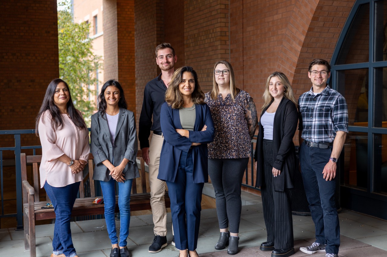
Members of UC Associate Professor Leyla Esfandiari's lab, from left, are Manju Sharma, Maulee Sheth, Maksym Krutko, Esfandiari, Holly Poling, Yara Izhiman and Gregory Paul Macke.
Related Stories
UC joins Ohio to improve worker safety
September 9, 2024
Ohio is taking steps to ensure the safety of workers in proximity to these electronic tools. The Bureau of Workers’ Compensation awarded $9.4 million for workforce safety innovation projects, including two led by UC’s College of Engineering and Applied Science.
What computers tell us about synthetic biology
March 3, 2022
Creating synthetic life could be easily within our grasp soon based on a comparison with the evolution of computer chips. Computer programming and gene synthesis appear to share little in common. But according to University of Cincinnati professor Andrew Steckl, an Ohio Eminent Scholar, leaps forward in technology in the former make him optimistic that wide scale gene manufacture is achievable.
Getting under your skin for better health
January 20, 2023
Biomedical engineers at the University of Cincinnati say interstitial fluid, the watery fluid found between and around cells, tissues or organs in the body, could provide an excellent medium for early disease diagnosis or long-term health monitoring.