Dual-Imaging Technique Useful Before-and During-Brain Surgery

CincinnatiBrain specialists associated with the Neuroscience Institute at the University of Cincinnati (UC) and University Hospital say the ability to incorporatein real timetwo high-tech imaging tools into the operating room can improve the functional abilities of patients who undergo brain surgery.
Neuroradiologist James Leach, MD, and his UC colleagues are among the first in the United States to combine functional magnetic resonance imaging (MRI) and diffusion tensor tractography data to outline important areas of brain function and their connections prior to surgeryand then transfer that data to the operating room to track those areas during actual surgery.
This approach, Leach says, has already been used in 20 cases at Cincinnatis University Hospital.
Combining tractography and functional MRI intraoperatively has been in use clinically for only a few years, says Leach, associate professor of radiology at UC and a neuroradiologist with University Hospital, but our preliminary data is very promising because it allows us to optimize surgical approaches to treating brain tumors to ultimately improve patient outcomes.
He says that combining standard visual, sound, and voice-based tests, whose results are measured by MRI, performed on a high-field-strength (3 Tesla) scanner, with a precise intraoperative guidance system will improve patients post-operative speech, movement and memory, and also optimize quality of life for those with brain tumors and other neurological conditions.
Leach will present this preliminary research in a special scientific exhibit at the American Society of Neuroradiologys annual meeting, June 913 in Chicago.
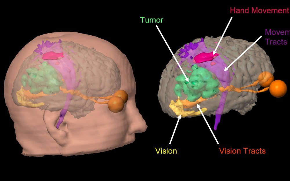
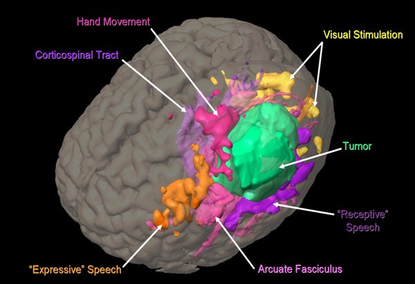
To combine both functional MRI and diffusion tensor tractography images in the operating room, Leach and his colleagues use a high-tech surgical navigation system known as BrainLAB. The BrainLAB technology incorporates functional MRI data to identify brain areas linked to speech, movement and vision, and diffusion tensor imaging data to map critical white-matter tractsthe electrical connections between different parts of the brain that should be avoided during surgery.
Leach says the detailed, easy-to-understand images produced from functional MRI data also help patients understand what is happening in their head and how the surgery will be conducted.
We can essentially show patients a color-coded picture of their brain and explain to them where their tumor is in relation to areas that control critical functions like expressive speech, movement and vision, says Leach. This can help alleviate the patients apprehension and help explain what will happen during surgery.
Although researchers expect this dual-imaging technique to become standard practice across the United States in the next five years, there are still limitations. For example, explains Leach, patients with limited vision may not demonstrate as much activity in the brain in areas linked to vision during functional MRI testing.
That doesnt necessarily mean that theres no function thereit may just be that functional MRI is not sensitive enough to detect it, says Leach.
Collaborators in this study include UC and Neuroscience Institute colleagues Christopher McPherson, MD, Ronald Warnick, MD, Raj Narayan, MD, and John Tew, MD.
Functional MRI helps neuroradiologists identify important areas of brain function.

James Leach, MD, specializes in brain imaging at UC and University Hospital.

Functional MRI helps neuroradiologists identify important areas of brain function.
Tags
Related Stories
UC study: Brain organ plays key role in adult neurogenesis
July 2, 2024
The University of Cincinnati has published research in the Proceedings of the National Academy of Sciences that found the choroid plexus and cerebrospinal fluid play a key role in maintaining a pool of newly born neurons to repair the adult brain after injury.
Put down that beer; it's not a tanning lotion
July 1, 2024
The University of Cincinnati's Kelly Dobos joined WVXU's Cincinnati Edition to discuss what's fact and what's myth when it comes to sunscreen use, different kinds of sunscreen and a social media recommendation to use beer on your skin to help get a tan.
Cincinnati researchers want to know if MRIs can work better
June 28, 2024
WVXU and the Cincinnati Business Courier highlighted a new collaboration between the University of Cincinnati College of Medicine, UC Health GE HealthCare, JobsOhio, REDI Cincinnati and Cincinnati Children’s to create an MRI Research and Development Center of Excellence located on UC’s medical campus.