Neuromuscular Center Able to Perform Fascicular Sciatic Nerve Biopsy

The ability to perform a delicate type of biopsy on major nerves is now one of the diagnostic tools available to specialists at the University of Cincinnati Neuroscience Institutes Neuromuscular Center. Hani Kushlaf, MD, a UC Health neurologist and an assistant professor of neurology and pathology, said the capability to perform a fascicular sciatic nerve biopsy is available at only a few elite academic medical centers in the United States.
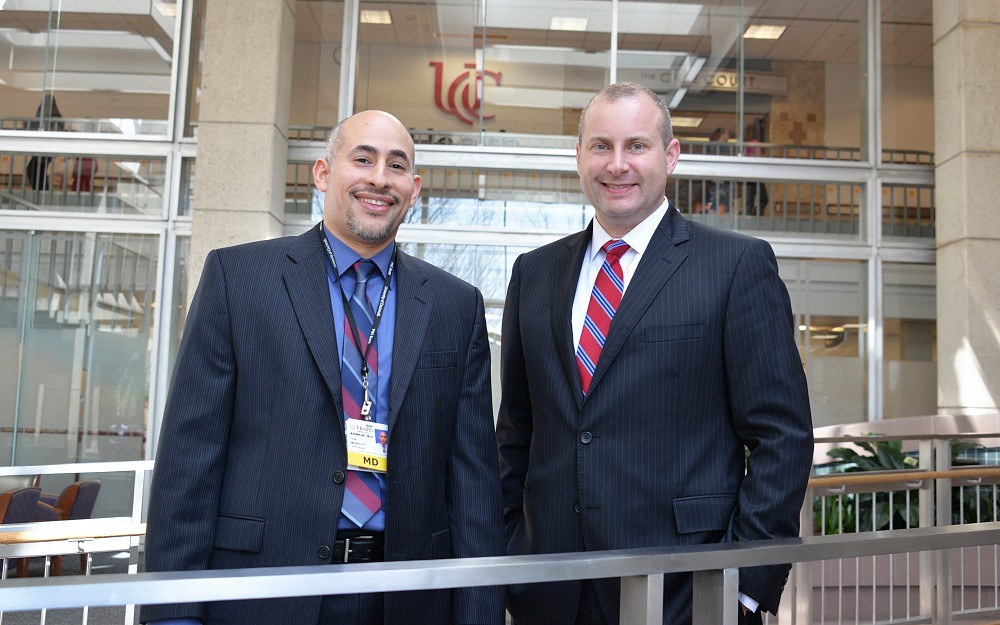
Kushlaf has teamed up with David Megee, MD, a UC Health plastic surgeon with microsurgical skills and special training in peripheral nerve surgery, to offer the outpatient diagnostic test to patients who have symptoms of a nerve disorder in a major nerve.
"This is new at UC Health, Kushlaf says. "It has not been done here before this year. Its important for hospitals and physicians in our region to know that we now have this diagnostic capability.
The fascicular nerve biopsy is used to diagnose conditions such as nerve vasculitis, neurolymphomatosis, sarcoidosis and benign or malignant nerve tumors. In the past, patients requiring the diagnostic procedure would have to travel out of town to institutions such as the Mayo Clinic or Johns Hopkins University.
A fascicular nerve biopsy differs from a standard nerve biopsy in important ways.
"During a typical nerve biopsy, we cut out a segment that represents the entire thickness of the nerve, Kushlaf says. That nerve is usually in a distal location, far from the center of the body. The sural nerve, which runs from the calf to the ankle, is a distal nerve that is often used in a standard nerve biopsy. The nerve is then examined under a microscope, and a diagnosis made.
In cases where a patients symptoms involve a proximal nervea major nerve that is near the center of the bodythe nerve cannot be cut, or major neurological deficits such as weakness or numbness would result. Proximal nerves include the sciatic nerve, which emanates from the lower back and runs down the back of the thigh.
To biopsy the sciatic nerve, Megee must first open the nerve to reveal the fascicles. Fascicles are bundles of nerve fibers, or axons, surrounded by a protective sheath. Fascicles look like small tubes running through a large tube (the nerve), and their job is to send electrical signals to the muscles and senses. If a healthy fascicle attached to a muscle is stimulated, it will cause the muscle to twitch.
Working under a microscope, Megee uses stimulating techniques to identify two or three non-functioning fascicles. He then removes them and sends them to Kushlaf, who doubles as a pathologist.
"We select non-functioning fascicles so that they show abnormalities under the microscope, Kushlaf says. "We want something involved in the disease process, something that will reveal abnormalities and allow us to make a diagnosis.
Fascicular sciatic nerve biopsy is the third major diagnostic tool that Kushlaf has helped bring to the Neuromuscular Center. The others are single-fiber EMG and peripheral ultrasound diagnostics. Kushlaf came to Cincinnati in 2012 after performing fellowships in muscle disorders, peripheral nerve disorders, and advanced neuromuscular medicine at the Mayo Clinic and Duke University.
Kushlaf and Megee are not expecting to do a multitude of fascicular sciatic nerve biopsy cases. "It is a rare procedure that is also applicable to the brachial plexus, lumbosacral plexus and major limb nerves, Kushlaf says, "but it is good to have it in our diagnostic armamentarium. When I was training at the Mayo Clinic, we saw two to three patients per week undergo this. Why? Because those patients were sent from other institutions.
"Patients from our region who were previously sent out of town can now rest assured that this diagnostic tool is now available here, at their own academic health center.
Tags
Related Stories
Don’t fall for these seven myths of research
July 14, 2025
A new publication by two University of Cincinnati researchers contends that adjusting how researchers approach their statistical analysis has the potential to change the lives of children and adolescents struggling with mental health issues across the world. Jeffrey Mills, PhD, and Jeffrey Strawn, MD, have been collaborating on interdisciplinary research and data analysis for years. Their latest paper, “Myths of Randomized Controlled Trial Analysis in Pediatric Psychopharmacology,” was selected as an Editor’s Pick for the Journal of Child and Adolescent Psychopharmacology.
Study finds pregnancy risks higher with ART in kidney transplant...
July 14, 2025
The odds of complications during pregnancy may be increased among women with kidney transplants conceiving through assisted reproductive technology (ART), according to a recent study led by the University of Cincinnati's Silvi Shah, MD, and published in Transplantation.
Just 30 minutes a day of ‘Japanese walking’ may help you get in...
July 14, 2025
The University of Cincinnati's Barbara Walker, PhD, was featured in a Washington Post article discussing interval walking training, or "Japanese walking," after the the technique has recently gone viral on TikTok.